The 19 Muscles Of The Foot - The medial and lateral pillars of the arch is formed by medial and lateral longitudinal arch respectively.. Plantar aponeurosis, and long & short plantar ligaments provide support for lla. When these bones are out of alignment, so is the rest of the body. There are 19 or 20 intrinsic foot muscles, therefore 38 to 40 intrinsic foot muscle tendons. Apex of the mla is superior articular surface of talus. Each toe has proximal and distal ip joints except for the great toe which only has one ip joint.
The distal tarsal rows including the three cuneiform bones and cuboid articulate with the base of each metatarsal to form the tmt complex. Only a small percentage of the population is born with foot problems. The common use of windlass is seen in pulling the anchor of the ship known as anchor windlass. Flexion of 4 lesser toes at metatarsophalangeal, proximal & distal interphalangeal joints inversion of foot plantar flexion of ankle. Later in the gait cycle, the foot then needs to act as a rigid lever to propel the weight of the body forward which is made possible by mt joint locking.
112 Human Foot Muscles 19 Century Medical Illustration Illustrations Clip Art Istock from media.istockphoto.com Chain reactions occur secondary to positioning of the foot. Muscles & tendon sheaths of the foot. Cote et al15concluded that postural stability is affected by foot position im both static and dynamic conditions. In addition to plantar aponeurosis the mla is also supported by spring ligament and deltoid ligament. The bony component of the arch consists of metatarsal heads, cuboids and 3 cuneiform bones. The medial and lateral pillars of the arch is formed by medial and lateral longitudinal arch respectively. Mobility is necessary for absorbing the ground reaction force of the body.12 subtalar pronation has a shock absorbing effect during initial heel contact.1211615 pronation is also necessary to make rotation of the leg possible and to absorb the impact of this rotation. It is a hinge joint and allows for dorsiflexion and plantarflexion movements in the sagittal plane.
Reduced or limited mobility in the lower limbs can be caused by a articular limitation.11615 in these cases some classic mobilizations or mobilizations according to manual therapy can be designated.11615 when the cause is a muscle shortening some stretching can be designated.
The interphalangeal joints of the toes are formed between the phalanges of the toes. Apex of the mla is superior articular surface of talus. The foot needs sufficient mobility to move into all the positions of the gait cycle while maintaining mobility and stability.812 physiological mobility is essential, because if mobility was too large, the foot would not have the capacity to be stable. The foot and ankle provide various important functions which includes: The deep peroneal nerve innervates all the muscles of the anterior compartment. There are 19 or 20 intrinsic foot muscles, therefore 38 to 40 intrinsic foot muscle tendons. Chondromalacia, shinsplints.) when there are some abnormalities in the normal gait cycle of functions of the body, some functional ortheses can be used.11615 this orthese have the capacity to correct the biomechanical function of the foot.11615 in contrast, insoles only support the arch of the foot. In addition to plantar aponeurosis the mla is also supported by spring ligament and deltoid ligament. It functions as a rigid structure for weight bearing and it can also function as a flexible structure to conform to uneven terrain. 1/4 of all the bones in the human body are down in your feet. During pronation/eversion of the foot, the axis of the tn and cc joints are parallel to each other, making it easier for them to independently move and unlock the mt joint. The transition from eversion to inversion is facilitated by the tibialis posterior muscle.12 the muscle is stretched like a spring and potential energy is stored.12 at the end of the midstance, the muscle passes from eccentric to concentric work and the energy is released. Tibialis anterior and posterior muscles play an important role in raising the medial border of the arch, whereas flexor hallucis longus acts as bowstring.
Lateral longitudinal arch (lla) it is the lowest arch and compromises of calcaneus, cuboid and fourth & fifth metatarsal as its bony component. In the transition from midstance to propulsion phase, the mechanisms often fail. This actions brings the metatarsal and tarsal bones together converting it into a rigid structure and eventually causing the longitudinal arches to rise. Jul 18, 2016 · muscle anatomy of the foot. The anterior compartment is comprised of four muscles that extend (dorsiflex) the foot and ankle (figure 14).
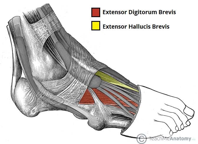
Muscles Of The Foot Dorsal Plantar Teachmeanatomy from teachmeanatomy.info Also, good (running) shoes are indicated.18 Subtalar pronation plays a role in shock absorption through eccentric control of the supinators.12 on the other side, the joint of chopart becomes unlocked so that the forefoot can stay loose and flexible.1 in midstance, the foot needs mobility to adapt to variation in surfaces.1211615 normal foot function provides the foot with the capacity to transform at the right time from a mobile adapter to a rigid lever. May 13, 2021 · medial and lateral tubercles of the calcaneum in a continuous line. It is also known as the talocalcaneal joint and is formed between the talus and calcaneus. Chain reactions occur secondary to positioning of the foot. Tibialis anterior and posterior muscles play an important role in raising the medial border of the arch, whereas flexor hallucis longus acts as bowstring. The medial and lateral pillars of the arch is formed by medial and lateral longitudinal arch respectively. The distal and inferior aspect of the tibia known as the plafond is connected to the fibula via tibiofibular ligaments forming a strong mortise which articulates with the talar dome distally.
The arches of foot provide functions of force absorption, base of support and acts as a rigid lever during gait propulsion.
Also known as lisfrancs joint. The tibialis anterior, the extensor hallucis longus, the extensor digitorum longus and the peroneus tertius. The deep peroneal nerve innervates all the muscles of the anterior compartment. Muscle layers of the sole of the foot. Plantar aponeurosis forms the supporting beam connecting the two pillars1. And during supination, mt joint locks providing rigidity of the foot and maximizing stability. It is also known as the talocalcaneal joint and is formed between the talus and calcaneus. This mechanism can be seen in foot. Jul 18, 2016 · muscle anatomy of the foot. In the transition from midstance to propulsion phase, the mechanisms often fail. The axis of the subtalar joint lies about 42o superiorly to the sagittal plane and about 16 to 23o medial to the transverse plane.89 the literature presents vast ranges of subtalar motion ranging from 5 to 65o.9 the average rom for pronation is 5o and 20o for supination. They include the abductor halluces, the flexor digitorum brevis, the abductor digiti minimi, and the quadratus plantae. The distal and inferior aspect of the tibia known as the plafond is connected to the fibula via tibiofibular ligaments forming a strong mortise which articulates with the talar dome distally.
The foot needs the capacity to bear body weight and act as a stable lever to propel the body in forward.1211615 this function requires pronation control of the subtalar joint.11615 Lateral longitudinal arch (lla) it is the lowest arch and compromises of calcaneus, cuboid and fourth & fifth metatarsal as its bony component. In addition to plantar aponeurosis the mla is also supported by spring ligament and deltoid ligament. Only a small percentage of the population is born with foot problems. Mobility is necessary for absorbing the ground reaction force of the body.12 subtalar pronation has a shock absorbing effect during initial heel contact.1211615 pronation is also necessary to make rotation of the leg possible and to absorb the impact of this rotation.
Foot Muscles Anatomy Part 1 2 Youtube from i.ytimg.com Blackwood et al11 concluded in the study that there is increased forefoot movement when the calcaneus is everted. In addition to plantar aponeurosis the mla is also supported by spring ligament and deltoid ligament. Whereas if the foot is stuck supinated, the midfoot would be hypomobile, which would compromise the ability of the foot to adjust to the terrain and increase demand on surrounding structures to maintain postural stability and balance. In the transition from midstance to propulsion phase, the mechanisms often fail. The foot contains 26 bones, 33 joints, 107 ligaments and 19 muscles. Muscle layers of the sole of the foot. The anterior compartment is comprised of four muscles that extend (dorsiflex) the foot and ankle (figure 14). The bony component of the arch consists of metatarsal heads, cuboids and 3 cuneiform bones.
The foot requires sufficient mobility and stability for all of its functions.
The distal and inferior aspect of the tibia known as the plafond is connected to the fibula via tibiofibular ligaments forming a strong mortise which articulates with the talar dome distally. The tip of the medial malleoli is anterior and superior to the lateral malleoli, which makes its axis oblique to both the sagittal and frontal planes. Also known as lisfrancs joint. The deep peroneal nerve innervates all the muscles of the anterior compartment. The foot is subdivided into the rearfoot, midfoot, and forefoot. And during supination, mt joint locks providing rigidity of the foot and maximizing stability. Windlass mechanism of foot the plantar aponeurosis acts similarly as windlass mechanism. Reduced or limited mobility in the lower limbs can be caused by a articular limitation.11615 in these cases some classic mobilizations or mobilizations according to manual therapy can be designated.11615 when the cause is a muscle shortening some stretching can be designated. They include the abductor halluces, the flexor digitorum brevis, the abductor digiti minimi, and the quadratus plantae. The common use of windlass is seen in pulling the anchor of the ship known as anchor windlass. Later in the gait cycle, the foot then needs to act as a rigid lever to propel the weight of the body forward which is made possible by mt joint locking. Whereas if the foot is stuck supinated, the midfoot would be hypomobile, which would compromise the ability of the foot to adjust to the terrain and increase demand on surrounding structures to maintain postural stability and balance. The axes cross each other during supination/inversion which locks the mt joint making it difficult to move.


0 Komentar